KPV is a naturally occurring tripeptide consisting of the amino acids
lysine, proline, and valine (Lys-Pro-Val), corresponding to positions 11-13
at the C-terminal end of alpha-melanocyte stimulating hormone (alpha-MSH).
Alpha-MSH is a 13-amino acid neuropeptide produced by post-translational
processing of proopiomelanocortin (POMC) in the pituitary gland, skin,
immune cells, and gut epithelium.
While alpha-MSH is well known for its role in melanogenesis and appetite
regulation through melanocortin receptors (particularly MC1R and MC4R),
research beginning in the 1980s and 1990s demonstrated that the C-terminal
tripeptide fragment KPV retains significant anti-inflammatory activity
despite being too small to bind to melanocortin receptors.
The anti-inflammatory properties of alpha-MSH were first recognized in
classical antipyretic assays, where the peptide reduced fever induced by
interleukin-1 and other pyrogens. Systematic structure-activity studies by
James Lipton, Anna Catania, and colleagues established that the minimal
anti-inflammatory sequence of alpha-MSH could be narrowed to the C-terminal
tripeptide KPV. This finding was surprising because it demonstrated that
anti-inflammatory signaling by alpha-MSH fragments could occur through a
mechanism entirely independent of the melanocortin receptor system.
KPV has subsequently attracted significant research interest as a minimal
anti-inflammatory peptide with potential therapeutic applications in
inflammatory conditions, particularly inflammatory bowel disease (IBD).
Its small size, oral bioavailability potential, and favorable safety
profile in preclinical models make it an attractive candidate for further
development.
The central anti-inflammatory mechanism of KPV involves inhibition of the
nuclear factor kappa-B (NF-kB) signaling pathway, the master
transcriptional regulator of inflammatory gene expression. NF-kB normally
resides in the cytoplasm in an inactive form, bound to its inhibitor
IkB-alpha. Upon stimulation by pro-inflammatory signals (such as TNF-alpha,
IL-1beta, or lipopolysaccharide), the IkB kinase (IKK) complex
phosphorylates IkB-alpha, targeting it for ubiquitin-mediated proteasomal
degradation. This releases the NF-kB p65/p50 heterodimer, which
translocates to the nucleus and activates transcription of inflammatory
genes including cytokines, chemokines, adhesion molecules, and inducible
enzymes.
KPV has been demonstrated to inhibit NF-kB activation at multiple points
in this cascade. Research by Ichiyama et al. (1999) showed that KPV
inhibits the activation of the IKK complex, thereby preventing
phosphorylation and degradation of IkB-alpha. As a result, NF-kB remains
sequestered in the cytoplasm and cannot activate inflammatory gene
transcription.
The downstream consequence is reduced expression of pro-inflammatory
cytokines including TNF-alpha, IL-6, IL-8, and IL-1beta, as well as
reduced expression of inducible nitric oxide synthase (iNOS) and
cyclooxygenase-2 (COX-2). This broad suppression of NF-kB-dependent
inflammatory mediators accounts for the wide-ranging anti-inflammatory
effects observed with KPV in multiple experimental systems.
A critical distinguishing feature of KPV is that its anti-inflammatory
activity does not require melanocortin receptor binding. Full-length
alpha-MSH exerts anti-inflammatory effects partly through MC1R activation
and partly through receptor-independent mechanisms. KPV, being only three
amino acids in length, is too small to engage melanocortin receptors with
meaningful affinity.
This receptor-independent mechanism was confirmed by studies showing that
KPV retains anti-inflammatory activity in cells lacking melanocortin
receptor expression and in the presence of melanocortin receptor
antagonists.
The receptor-independent mechanism involves direct intracellular action.
KPV has been shown to cross cell membranes and accumulate within the cell,
where it can directly interact with components of the NF-kB signaling
pathway. Studies have demonstrated that KPV can even enter the cell
nucleus, where it may directly interfere with NF-kB binding to DNA
promoter elements. This intracellular mode of action distinguishes KPV
from most peptide therapeutics, which typically act at cell surface
receptors.
A key finding with significant implications for the therapeutic development
of KPV was the discovery that it is a substrate for the intestinal peptide
transporter PepT1 (SLC15A1). PepT1 is a proton-coupled oligopeptide
transporter expressed on the apical membrane of intestinal epithelial cells
that is responsible for the absorption of dietary di- and tripeptides from
the intestinal lumen.
Dalmasso et al. (2008) demonstrated that KPV is efficiently transported
across intestinal epithelial cells by PepT1, enabling its uptake from the
intestinal lumen into enterocytes. Importantly, PepT1 expression is
upregulated in the inflamed intestinal epithelium of patients with IBD,
potentially creating a natural targeting mechanism whereby more KPV is
absorbed at sites of intestinal inflammation.
Once inside epithelial cells, KPV can exert its anti-inflammatory effects
by inhibiting NF-kB and reducing production of inflammatory mediators.
This PepT1-mediated uptake mechanism provides a rationale for oral
administration of KPV for intestinal inflammatory conditions, as the
peptide can be absorbed directly into the cells most affected by mucosal
inflammation without requiring systemic absorption and distribution.
The most developed preclinical evidence for KPV pertains to its efficacy
in experimental models of inflammatory bowel disease. In the dextran sodium
sulfate (DSS) model of colitis, which mimics features of ulcerative colitis,
oral administration of KPV significantly reduced disease activity index
scores, colonic weight-to-length ratio (a measure of edema and
inflammation), and histological damage scores.
These benefits were associated with reduced tissue levels of TNF-alpha,
IL-6, and other pro-inflammatory cytokines, as well as reduced neutrophil
infiltration as measured by myeloperoxidase activity.
In the trinitrobenzene sulfonic acid (TNBS) model, which generates a
Th1-dominant inflammatory response more similar to Crohn's disease, KPV
similarly demonstrated protective effects, reducing mucosal damage,
inflammatory cell infiltration, and pro-inflammatory cytokine production.
These findings across mechanistically distinct colitis models suggest that
KPV's anti-inflammatory effects are relevant to both major forms of IBD.
Nanoparticle Delivery Studies#
To further enhance intestinal targeting and stability, researchers have
investigated encapsulation of KPV in nanoparticle delivery systems. Laroui
et al. (2010) developed alginate-chitosan nanoparticles loaded with KPV
and demonstrated that this formulation improved colonic delivery and
enhanced therapeutic efficacy in the DSS colitis model compared to free
KPV.
The nanoparticles protected KPV from luminal degradation, concentrated the
peptide at sites of inflammation through electrostatic interactions with
the inflamed mucosa, and enabled sustained release within the colonic
tissue. Hyaluronic acid-functionalized nanoparticles have also been
investigated, exploiting the overexpression of CD44 receptors on inflamed
colonic epithelium to achieve targeted delivery.
KPV has been investigated for anti-inflammatory effects in dermatological
contexts, consistent with the known expression and function of alpha-MSH
in the skin. In preclinical models of cutaneous inflammation, KPV reduced
inflammatory cell infiltration, edema, and expression of inflammatory
mediators.
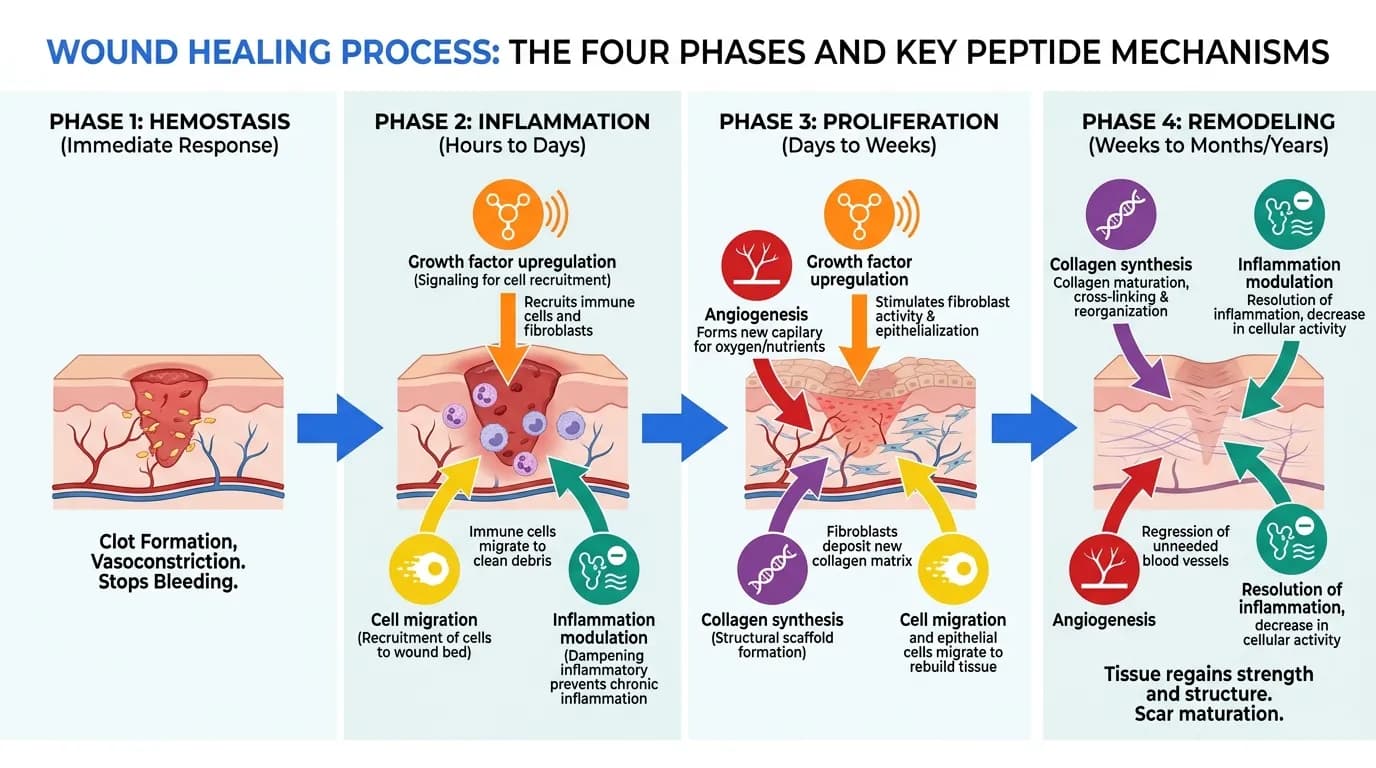
In wound healing models, KPV treatment has been associated with reduced
inflammatory phase duration and accelerated transition to the proliferative
phase, resulting in faster wound closure. However, dermatological
applications of KPV remain less developed than gastrointestinal
applications.
The primary therapeutic interest in KPV centers on inflammatory bowel
disease, where its oral bioavailability through PepT1 transport,
anti-inflammatory potency, and preferential uptake at inflamed mucosal
sites create a compelling therapeutic profile. Both ulcerative colitis and
Crohn's disease represent potential target conditions, based on efficacy
in DSS and TNBS models respectively.
Beyond IBD, KPV is under investigation for other conditions characterized
by mucosal inflammation, including pouchitis (inflammation of the ileal
pouch following colectomy for ulcerative colitis) and radiation proctitis.
The gut-targeted activity of KPV through PepT1 transport makes it
particularly suitable for conditions affecting the intestinal mucosa.
Systemic anti-inflammatory applications are also being explored, though
these would likely require parenteral administration or advanced oral
delivery systems to achieve adequate systemic bioavailability. Potential
systemic applications include rheumatoid arthritis, psoriasis, and other
chronic inflammatory conditions.
KPV is also being investigated as a component of combination therapies
with conventional IBD medications, including mesalamine, corticosteroids,
and biologics. The distinct mechanism of action (direct NF-kB inhibition
without immunosuppression) may provide additive or synergistic benefits
when combined with existing treatments.
Despite promising preclinical data, KPV has not yet been evaluated in
human clinical trials for any indication. The entirety of the evidence
base consists of preclinical studies in cell culture systems and animal
models. The translation of preclinical efficacy in rodent colitis models
to human IBD is historically challenging, as many agents showing strong
preclinical activity have failed to demonstrate meaningful clinical
benefits in human trials.
The pharmacokinetics of KPV in humans are not characterized. While
PepT1-mediated transport provides a mechanism for intestinal absorption,
the fraction of an oral dose that reaches the colonic epithelium (versus
absorption in the small intestine or degradation in the gastrointestinal
lumen) is uncertain. The stability of KPV in gastric acid and against
luminal proteases has not been fully characterized under physiological
conditions.
The precise intracellular molecular targets of KPV remain incompletely
defined. While NF-kB inhibition is well documented, the specific protein
interactions through which a three-amino-acid peptide achieves this
inhibition have not been definitively identified. Understanding these
interactions at the molecular level would facilitate rational optimization
of KPV analogs with improved potency or selectivity.
Long-term safety data are absent. While KPV is a naturally occurring
fragment of an endogenous peptide, chronic administration of
supraphysiological doses could theoretically produce unanticipated effects.
The consequences of sustained NF-kB inhibition in the intestinal
epithelium, where NF-kB signaling also plays roles in epithelial
homeostasis, barrier function, and antimicrobial defense, would need to
be carefully evaluated.
KPV's anti-inflammatory mechanism involves inhibition of NF-kB rather
than immunosuppression, which may be advantageous for safety but could
also limit efficacy in conditions requiring more profound
immunomodulation. The relative potency of KPV compared to established
anti-inflammatory biologics (anti-TNF antibodies, anti-integrin
antibodies, JAK inhibitors) in the context of human IBD is entirely
unknown.
Finally, intellectual property and commercial development challenges may
affect the translational trajectory of KPV. As a naturally occurring
tripeptide, KPV itself may face patentability challenges, which could
limit commercial interest in funding clinical trials. Proprietary
formulations and delivery systems (such as nanoparticle formulations)
represent one strategy for addressing this limitation.
PepT1-mediated tripeptide KPV uptake reduces intestinal inflammation, published in Gastroenterology (Dalmasso G et al., 2008; PMID: 18061177):
Demonstrated that KPV is a substrate for the intestinal peptide transporter PepT1, enabling its uptake from the intestinal lumen into epithelial cells where it inhibits NF-kB signaling and reduces inflammatory responses.
- KPV is efficiently transported by PepT1 across intestinal epithelial cell monolayers
- PepT1-mediated KPV uptake reduces NF-kB activation in colonocytes
- PepT1 expression is upregulated in inflamed intestinal epithelium
Silencing of peptide transporter PepT1 reduces colitis in mice by targeting epithelial NF-kB signaling, published in Gastroenterology (Dalmasso G et al., 2010):
Further characterized the role of PepT1 in KPV-mediated anti-inflammatory effects, demonstrating that PepT1 silencing abolishes KPV's protective effects in colitis models.
- PepT1 silencing blocked KPV uptake and anti-inflammatory activity
- Confirmed PepT1 as the essential transporter for KPV's intestinal effects
- Reinforced the NF-kB pathway as the downstream target
Functional nanoparticles for oral drug delivery of KPV peptide for colitis treatment, published in Biomaterials (Laroui H et al., 2010; PMID: 19909746):
Developed alginate-chitosan nanoparticles loaded with KPV for targeted colonic delivery, demonstrating improved therapeutic efficacy compared to free KPV in the DSS colitis model.
- Alginate-chitosan nanoparticles successfully encapsulated and protected KPV
- Nanoparticle formulation improved colonic delivery of KPV
- Enhanced therapeutic efficacy in DSS colitis model compared to free KPV
Anti-inflammatory effects of alpha-MSH C-terminal peptide KPV, published in Peptides (Ichiyama T et al., 1999):
Established that KPV inhibits the IKK complex, preventing IkB-alpha phosphorylation and NF-kB nuclear translocation, thereby suppressing inflammatory gene expression.
- KPV inhibits IKK complex activation
- Prevents phosphorylation and degradation of IkB-alpha
- Blocks NF-kB nuclear translocation