IGF-1 LR3 vs MGF: Growth Factor Peptides for Muscle Research
IGF-1 LR3 vs MGF compared for muscle growth research — mechanisms, satellite cell effects, half-life differences, and evidence levels.
Verdict at a Glance
IGF-1 LR3 and MGF represent two distinct phases of the IGF-1 system in muscle biology. MGF is the endogenous initiator of satellite cell proliferation following mechanical damage, while IGF-1 LR3 is an engineered analog providing potent, sustained IGF-1 receptor activation for differentiation and growth. Neither has human clinical trial data, keeping both in the preclinical research domain. IGF-1 LR3 has broader practical utility due to its established role in cell culture and longer half-life, while MGF offers unique insight into the early repair signaling cascade.
| Best for | Pick | Why |
|---|---|---|
| Cell culture and bioprocessing applications | IGF-1 LR3 | Industry-standard growth factor supplement with well-characterized potency and extended bioavailability in culture media |
| Satellite cell activation and muscle repair research | MGF | Specifically activates quiescent satellite cells through a mechanism distinct from IGF-1 receptor signaling |
| Sustained growth factor signaling in research | IGF-1 LR3 | Reduced IGFBP binding provides 2-3x longer half-life and greater effective potency than native IGF-1 |
| Understanding exercise-induced muscle repair mechanisms | MGF | Endogenous expression pattern after mechanical loading makes it the physiologically relevant repair initiator |
| Category | IGF-1 LR3 | MGF | Advantage |
|---|---|---|---|
| Mechanism of Action | Modified IGF-1 with dramatically reduced IGFBP binding; activates the IGF-1 receptor to drive PI3K/AKT and MAPK pathways for cell proliferation, differentiation, and protein synthesis | 24-amino acid E-domain peptide from IGF-1Ec splice variant; activates satellite cells through IGF-1R-independent signaling; initiates proliferation phase before differentiation | Comparable |
| Research Evidence | Extensively used in cell culture and bioprocessing; well-characterized in vitro potency; limited in vivo clinical data in humans | Primarily preclinical evidence from rodent and cell culture models; discovered in the late 1990s; no human clinical trials | IGF-1 LR3 |
| Side Effect Profile | Potential for hypoglycemia due to insulin receptor cross-reactivity; theoretical oncogenic risk from potent growth factor signaling; no approved safety database | Minimal reported adverse effects in preclinical studies; rapid degradation limits systemic exposure; no human safety data available | Comparable |
| Dosing Complexity | Extended half-life due to reduced IGFBP binding simplifies dosing frequency; used at microgram levels in research; requires careful handling | Very short half-life (minutes) requires frequent administration or PEG modification; local injection protocols studied in preclinical models | IGF-1 LR3 |
| Biological Role | Engineered analog optimized for maximum IGF-1 receptor activation; promotes both proliferation and differentiation of myoblasts and other cell types | Endogenous repair factor specifically expressed after mechanical damage; initiates satellite cell activation as the first phase of muscle repair cascade | Comparable |

Introduction#
IGF-1 LR3 and MGF are both products of the insulin-like growth factor-1 system, yet they serve fundamentally different biological roles. IGF-1 LR3 is an engineered analog of mature IGF-1, designed to maximize receptor activation by escaping the binding proteins that normally sequester circulating IGF-1. MGF (Mechano Growth Factor) is the unique E-domain peptide from the IGF-1Ec splice variant, a naturally occurring repair signal expressed locally in response to mechanical tissue damage.
Understanding where each peptide fits within the IGF-1 signaling cascade is essential for appreciating their distinct research applications and biological significance.
Quick Comparison#
| Feature | IGF-1 LR3 | MGF |
|---|---|---|
| Structure | 83 amino acids, 9.1 kDa | 24 amino acids, 2.9 kDa |
| Origin | Engineered IGF-1 analog | Endogenous IGF-1Ec splice variant E-domain |
| Receptor | IGF-1 receptor (IGF-1R) | IGF-1R-independent mechanism |
| Primary Role | Cell proliferation and differentiation | Satellite cell activation (repair initiation) |
| IGFBP Binding | Dramatically reduced | Not applicable |
| Half-Life | Extended (hours) | Very short (minutes) |
| Research Status | Preclinical | Preclinical |
| Primary Use | Cell culture, bioprocessing | Muscle repair research |
| PEGylated Form | Not standard | PEG-MGF available |
Mechanism of Action Comparison#
IGF-1 LR3#
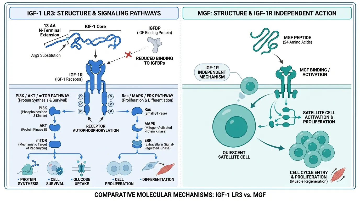
IGF-1 LR3 is an 83-amino acid analog of native IGF-1 incorporating two structural modifications: an arginine substitution at position 3 and a 13-amino acid N-terminal extension. These changes reduce binding to IGF binding proteins (IGFBPs) by approximately 100-fold while preserving full affinity for the IGF-1 receptor (IGF-1R).
Upon binding IGF-1R, a receptor tyrosine kinase, IGF-1 LR3 triggers autophosphorylation of the receptor's intracellular beta subunits. This initiates two major signaling cascades:
- PI3K/AKT/mTOR pathway: Drives protein synthesis, cell survival, and glucose uptake through IRS-1/2 adaptor proteins. mTOR activation downstream promotes ribosomal biogenesis and translation initiation.
- Ras/MAPK/ERK pathway: Promotes cell proliferation, differentiation, and gene expression changes through nuclear transcription factor activation.
Because approximately 98% of native IGF-1 is normally sequestered by IGFBPs, the reduced binding of IGF-1 LR3 results in dramatically higher effective free concentrations. This makes it 2-3 times more potent than native IGF-1 in most cell-based assays and the preferred form for cell culture applications.
MGF#
MGF is the 24-amino acid C-terminal E-domain peptide unique to the IGF-1Ec splice variant (IGF-1Eb in rodents). Unlike IGF-1 LR3 which activates the canonical IGF-1 receptor, MGF appears to have independent biological activity that does not require IGF-1R signaling.
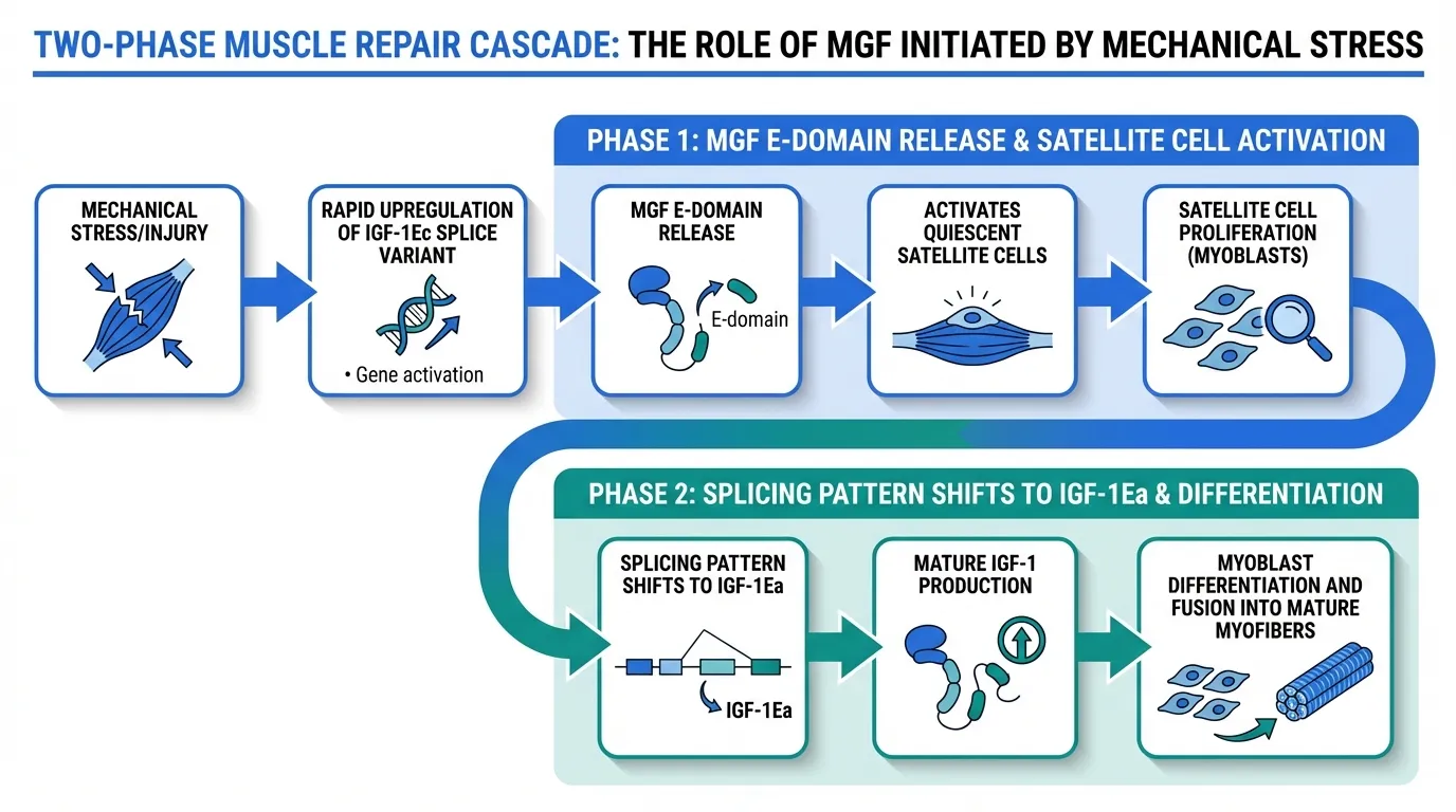
The primary biological role of MGF involves activating quiescent satellite cells, the resident stem cells in skeletal muscle. Following mechanical stress or injury, MGF expression is temporally regulated as the first phase of a two-phase repair cascade:
- Phase 1 (MGF): The IGF-1Ec splice variant is rapidly upregulated after damage. The MGF E-domain activates quiescent satellite cells, stimulating their entry into the cell cycle and proliferation as myoblasts.
- Phase 2 (IGF-1Ea): The splicing pattern shifts to favor IGF-1Ea, producing mature IGF-1 that drives differentiation and fusion of the expanded myoblast pool into mature myofibers.
This temporal sequence positions MGF as the initiating signal for repair, responsible for expanding the precursor cell pool before the differentiation signals encoded by mature IGF-1 take over. The mechanism may involve distinct receptors or signaling intermediates not shared with IGF-1R activation.
Evidence and Research Comparison#
IGF-1 LR3 Research#
IGF-1 LR3 has become the standard form of IGF-1 used in laboratory research and biomanufacturing:
- Cell culture: Widely used as a growth factor supplement in serum-free and low-serum culture media, where it provides significantly greater potency per unit mass compared to native IGF-1
- Bioprocessing: Applied in manufacturing of biopharmaceuticals and cell-based therapies to support cell growth and viability
- In vitro pharmacology: Extensive characterization of receptor binding kinetics, downstream signaling, and effects on various cell types including myoblasts, osteoblasts, and neural progenitors
- Animal studies: Some preclinical data on systemic and local administration effects on muscle growth and recovery in rodent models
No human clinical trials have been conducted with IGF-1 LR3. Its pharmacological profile is inferred from in vitro work and from studies of native IGF-1 and the FDA-approved recombinant IGF-1 (mecasermin/Increlex).
MGF Research#
MGF research originates primarily from the laboratory of Geoffrey Goldspink at University College London:
- Discovery and characterization: First described in the late 1990s as a mechanically sensitive IGF-1 splice variant in rabbit and human skeletal muscle
- Satellite cell activation: Demonstrated ability to activate quiescent satellite cells in culture, with evidence for IGF-1R-independent signaling
- Cardiac applications: Expression of IGF-1Ec documented in cardiac muscle following myocardial infarction, with preclinical evidence for cardioprotective effects
- Neuroprotection: MGF expression detected in brain tissue following ischemic injury, with preliminary evidence for neuronal survival effects
- Aging research: Impaired MGF upregulation documented in elderly subjects after exercise, suggesting a mechanism for age-related decline in regenerative capacity
MGF remains entirely preclinical, with no human clinical trials. Its rapid degradation in vivo (half-life of minutes) has been a practical limitation, addressed partly by PEGylated formulations (PEG-MGF).
Side Effects and Safety Comparison#
IGF-1 LR3 Side Effects#
No human safety data exist for IGF-1 LR3 specifically. Theoretical risks are extrapolated from IGF-1 biology:
- Hypoglycemia: IGF-1 shares structural homology with insulin and can activate insulin receptors, particularly at high concentrations
- Growth factor signaling: Sustained IGF-1R activation carries theoretical oncogenic potential, as the IGF-1 axis promotes cell proliferation and survival
- Systemic effects: Reduced IGFBP binding means greater systemic exposure to active growth factor, potentially amplifying both therapeutic and adverse effects
- Organ-specific: Potential for cardiac hypertrophy and other tissue growth with chronic exposure
MGF Side Effects#
MGF safety data are limited to preclinical observations:
- Rapid clearance: The very short half-life limits systemic exposure, which may reduce off-target effects
- Local action: Endogenous MGF acts primarily as a local paracrine factor, suggesting that targeted administration may have a favorable safety profile
- No human data: Without clinical trials, the safety profile in humans remains undefined
- Proliferative concerns: As a satellite cell activator, theoretical concerns exist regarding uncontrolled proliferation, though preclinical data have not demonstrated this
Dosing and Administration Comparison#
IGF-1 LR3 Dosing#
| Parameter | Details |
|---|---|
| Route | Subcutaneous or intramuscular in research |
| Research doses | 20-100 mcg per administration |
| Cell culture | 50-100 ng/mL in culture media |
| Frequency | Daily in research protocols |
| Half-life | Extended (hours, due to reduced IGFBP binding) |
| Storage | -20 C long-term; 4 C short-term after reconstitution |
MGF Dosing#
| Parameter | Details |
|---|---|
| Route | Local intramuscular injection in research |
| Research doses | 100-200 mcg per administration |
| Frequency | Daily or post-exercise in research |
| Half-life | Minutes (native form) |
| PEG-MGF | Extended half-life with PEGylation |
| Storage | -20 C long-term; reconstitute fresh |
The short half-life of native MGF is a significant practical limitation. PEG-MGF addresses this through polyethylene glycol conjugation, extending the half-life to hours and enabling less frequent administration in research settings.
Use Case Recommendations#
Choose IGF-1 LR3 When:#
- Cell culture and bioprocessing require a potent, cost-effective growth factor
- Sustained IGF-1R activation is the experimental objective
- Differentiation and protein synthesis pathways are the primary research targets
- Practical convenience is important, given the longer half-life
- Broad tissue effects beyond muscle are relevant to the study design
Choose MGF When:#
- Satellite cell biology and stem cell activation are the specific research focus
- Exercise-induced repair mechanisms are being studied
- Temporal signaling in muscle regeneration is the question being addressed
- IGF-1R-independent pathways in tissue repair are of interest
- Age-related decline in regenerative capacity is the model under investigation
Can They Be Combined?#
Combining IGF-1 LR3 and MGF in research models has biological rationale, as they represent sequential phases of the endogenous repair cascade. MGF initiates satellite cell proliferation, while IGF-1 LR3 could then drive differentiation and protein synthesis in the expanded cell pool. Some preclinical studies have explored sequential administration protocols designed to mimic this temporal pattern.
However, no standardized combination protocols exist, and the interaction between exogenous MGF and IGF-1 LR3 has not been systematically studied in vivo. Researchers exploring this approach should consider the vastly different half-lives and the potential for overlapping signaling to disrupt the endogenous temporal cascade.
Verdict#
IGF-1 LR3 and MGF occupy distinct positions within the IGF-1 signaling system. IGF-1 LR3 is the practical workhorse: an engineered analog with enhanced potency and stability that has become the standard growth factor for cell culture and in vitro research. MGF is the biological discovery: an endogenous repair initiator that reveals how tissues coordinate the early stages of regeneration following mechanical damage.
Neither peptide has progressed to human clinical trials, keeping both firmly in the preclinical research domain. For researchers working with established growth factor protocols, IGF-1 LR3 offers greater convenience and broader applicability. For those investigating the fundamental biology of muscle repair, satellite cell activation, and age-related regenerative decline, MGF provides unique mechanistic insight that IGF-1 analogs cannot replicate.
For related growth factor peptides, see our profiles on HGH 191AA and PEG-MGF, or explore the dosing calculator for weight-based research dose estimation.
Further Reading#

Which Is Better For...
Cell culture and bioprocessing applications
IGF-1 LR3
Industry-standard growth factor supplement with well-characterized potency and extended bioavailability in culture media
Satellite cell activation and muscle repair research
MGF
Specifically activates quiescent satellite cells through a mechanism distinct from IGF-1 receptor signaling
Sustained growth factor signaling in research
IGF-1 LR3
Reduced IGFBP binding provides 2-3x longer half-life and greater effective potency than native IGF-1
Understanding exercise-induced muscle repair mechanisms
MGF
Endogenous expression pattern after mechanical loading makes it the physiologically relevant repair initiator
Continue Your Research
Get comparison updates
We publish new head-to-head comparisons regularly. Subscribe to see them first.
Frequently Asked Questions About IGF-1 LR3 vs MGF: Growth Factor Peptides for Muscle Research
Which is better, IGF-1 LR3 or MGF?
IGF-1 LR3 and MGF represent two distinct phases of the IGF-1 system in muscle biology. MGF is the endogenous initiator of satellite cell proliferation following mechanical damage, while IGF-1 LR3 is an engineered analog providing potent, sustained IGF-1 receptor activation for differentiation and growth. Neither has human clinical trial data, keeping both in the preclinical research domain. IGF-1 LR3 has broader practical utility due to its established role in cell culture and longer half-lif... Individual responses may vary, and this comparison is based on available research data, not a treatment recommendation.
What are the key differences between IGF-1 LR3 and MGF?
The main differences across comparison categories are: Mechanism of Action: advantage goes to neither (tie); Research Evidence: advantage goes to IGF-1 LR3; Side Effect Profile: advantage goes to neither (tie). 2 additional categories are analyzed in the full comparison. Each category evaluates a different dimension of the two peptides.
When should I consider IGF-1 LR3 over MGF?
For the scenario of "Cell culture and bioprocessing applications," research data suggests IGF-1 LR3 may be more relevant. Industry-standard growth factor supplement with well-characterized potency and extended bioavailability in culture media. This is based on currently available evidence and individual circumstances may differ.
How do IGF-1 LR3 and MGF differ in their mechanisms of action?
IGF-1 LR3: Modified IGF-1 with dramatically reduced IGFBP binding; activates the IGF-1 receptor to drive PI3K/AKT and MAPK pathways for cell proliferation, differentiation, and protein synthesis. MGF: 24-amino acid E-domain peptide from IGF-1Ec splice variant; activates satellite cells through IGF-1R-independent signaling; initiates proliferation phase before differentiation.
Which has fewer side effects, IGF-1 LR3 or MGF?
In terms of side effects and tolerability, the advantage goes to neither (comparable). IGF-1 LR3: Potential for hypoglycemia due to insulin receptor cross-reactivity; theoretical oncogenic risk from potent growth factor signaling; no approved sa.... MGF: Minimal reported adverse effects in preclinical studies; rapid degradation limits systemic exposure; no human safety data available.
Medical Disclaimer
This website is for educational and informational purposes only. The information provided is not intended to diagnose, treat, cure, or prevent any disease. Always consult with a qualified healthcare professional before using any peptide or supplement.